Where’s the edge of the visual field

and what do cones have to do with it?

Vaguely summarized here and properly described in the paper:

  • precise and reliable measurement of the edge of the visual field
  • little influence of contrast on the result
  • using a colored stimulus, cones are demonstrated to be functional in this region

P Veto, PBM Thomas, P Alexander, T Wemyss and JD Mollon (2020)
‘The last channel’: vision at the temporal margin of the field
Proceedings of the Royal Society B, 287, 20200607

It’s common knowledge that central vision is dominated by cones (receptors to detect colors, packed for high resolution; also, they don’t work well in dim conditions) and peripheral vision is dominated by rods (no color information; crucial when it’s dark). Plus, the periphery aids in navigation and warns to sudden movement.

rod_and_cone_distribution

Indeed, motion perception is the last function that we still have at the edge of the visual field. You’ve probably experienced this e.g. while playing ball games or when an adjacent car on the highway did something unexpected.


But here’s the discrepancy: rods are slower than cones

– so why would they be the sentinel?


Conveniently (and much less commonly known), the very edge of the retina has a high relative density of cones. Their function has long been elusive, but here are a couple of hypotheses:

  1. Detecting sudden changes
  2. Measuring self-motion
  3. Contributing to color constancy
  4. Contributing to circadian rhythms

While the first two functions require some imaging of the external world, the latter two assume that light doesn’t even (have to) reach these cones directly. So far it has neither been fully clear if light directly projects to these cones, nor has there been clear evidence to suggest that they have any real function.

Here, we have made a couple of measurements to learn something new: cones actually appear to be responsible for motion perception at the far edge of the field.

First, we showed that by using a direction discrimination paradigm, the edge of the field can be measured reliably…

Test-retest reliability of the mean threshold eccentricity.

… and without much dependence on contrast (when that is reasonably high).

Change in threshold eccentricity with stimulus contrast.

Then, by replacing the black-and-white stimulus with a colored one, which is primarily visible to cones and not rods, we also found that the edge of the field remained at the same location. This could not be possible, if we used rods to solve the task.

Threshold eccentricity with black-and-white (control) and colored (silent substitution) stimuli.


Finally, an open question that we didn’t discuss much in the article

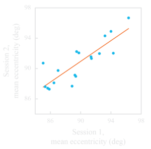
Pupil size did not correlate with the measured eccentricity of the visual field’s edge.where_is_the_edge_of_our_visual_field_and_what_do_we_see_there_2

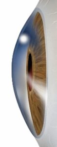
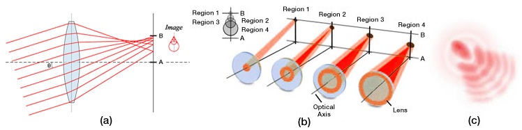
On the one hand, much less light can enter the eye from extreme angles (about 1/5th in this case); therefore, we could expect worse performance with smaller pupil diameter.

(Viewing angle in this illustration to the right is less than 90°. Due to refraction on the cornea however, the pupil would appear similar to this also from a 90° angle – do check on the person you next encounter.)

On the other hand, optical aberrations may take place and lead to worse imaging when the pupil is larger. This is due to coma and the “cat’s eye effect.”

Relationship between pupil size and comatic aberration.

It might be that we don’t see either of these effects in our data because

  • the two (positive & negative) cancel each other out
  • neither of them is significant enough to matter
  • they would only affect perception of motion in a certain direction (and not the one we tested; this refers to the optical aberrations only)

Here I just speculate of course, let me know if you have a better idea.